Skip to content

Imaging studies
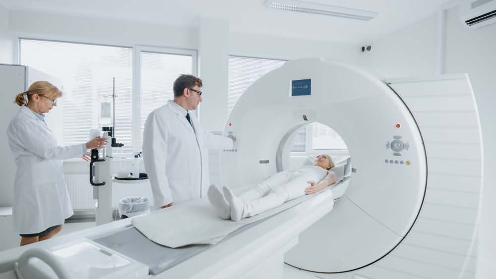
Those lab tests may be complemented with other tests and imaging studies to confirm the diagnosis or to detect the extent of damage to different body systems. The most commonly done imaging studies include:
- Xray: Xray imaging is needed in children with leg bowing, to confirm the diagnosis of rickets.
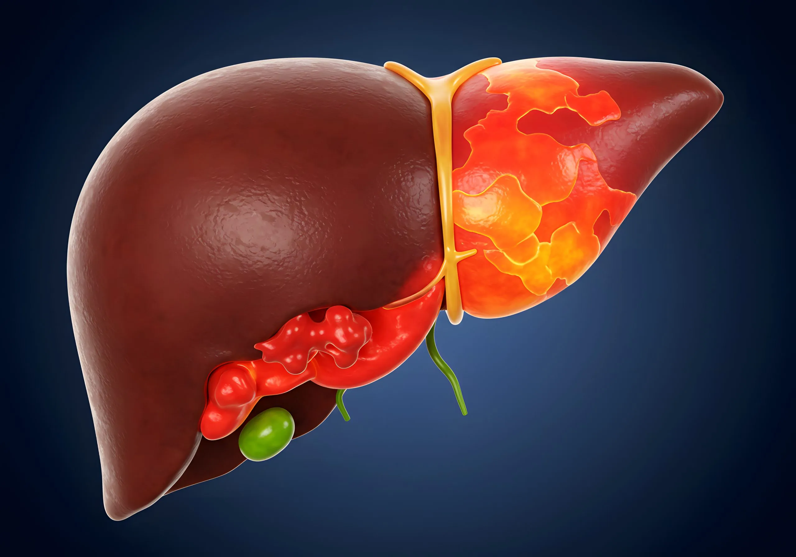
- Abdominal ultrasound: Abdominal ultrasound can assess the condition of the liver and kidneys, two organs commonly affected by Alagille syndrome.
- Echocardiography: Echocardiography is a special kind of ultrasound for the heart and is done in patients with Alagille syndrome. It is used to detect defects in the heart and problems with the heart valves.
- CT scan: CT scanning is a special type of Xray that is used to get a cross-section of the body and is used in Alagille to detect problems of the blood vessels whether the aorta, blood vessels of the brain or the kidney.
- Doppler scan: Doppler scan is a type of ultrasound that can detect the flow of blood and its pattern within blood vessels and is also used to detect narrowing of blood vessels of the kidney.
- OCT: Ocular coherent tomography is a type of scanning for the retina of the eye for detachment or swelling of the optic disc.
Even after all of the above investigations -with the exception of genetic analysis- there are several conditions that are similar to Alagille syndrome including all the causes of jaundice in children and the newborn, cystic fibrosis, and biliary atresia.