Skip to content

How is hemangioma diagnosed?

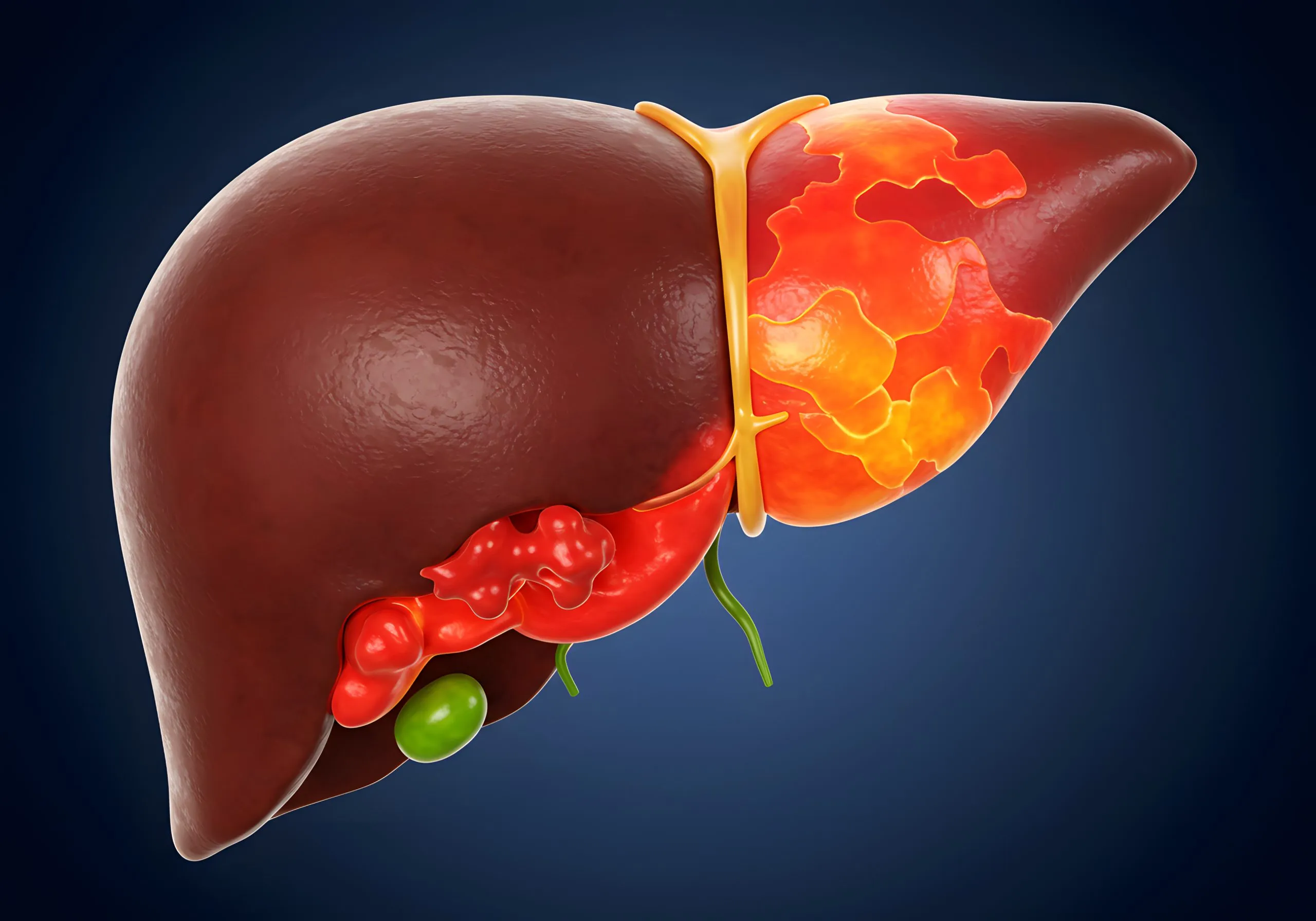
Hemangiomas are symptomless but can cause problems if they are a part of another disease or “syndrome”. A syndrome is a group of well-known symptoms and findings that usually result from a single disease process. Liver hemagiomas can occur in cases of systemic lupus of Klippel- Trenaunay syndrome. Otherwise, the main complaint is upper abdominal pain especially on the right side and a feeling of fullness. Your doctor will examine your abdomen and sometimes listen using their stethoscope because large hemangiomas produce a “murmur”.
Afterwards and to confirm the diagnosis, some lab tests and imaging studies are done. Lab tests such as a complete blood picture are routine. However, they can indicate a common complication of hemangiomas where platelets decrease in number. Also, liver function tests may be needed in some cases of large hemangiomas.
Imaging studies include a liver ultrasound, which can reveal the hemangioma, albeit with a low accuracy and it can be mistaken for any other benign or even malignant tumors. The main imaging study to confirm a hemangioma is a triphasic CT, which involves a CT scan of the liver in 3 stages with contrast injection. An MRI can be done in some cases when CT can’t be done as in pregnancy when hemangioma is diagnosed most. A biopsy is impossible in a hemangioma due to the risk of hemorrhage.