
Anatomic and histologic differences
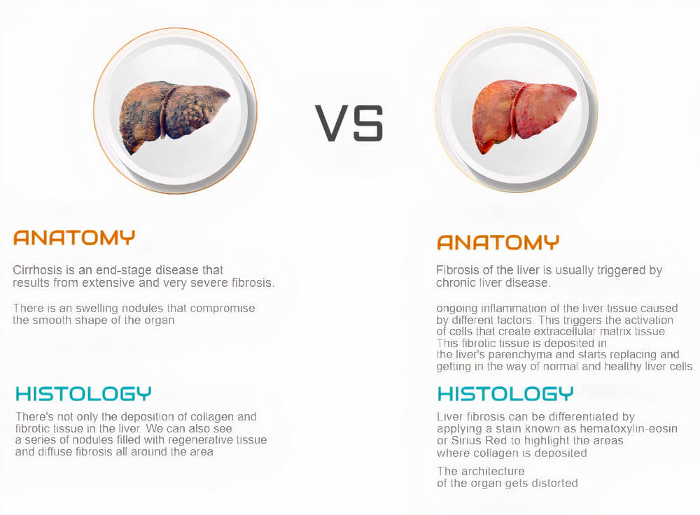
Fibrosis of the liver is usually triggered by chronic liver disease. There is ongoing inflammation of the liver tissue caused by different factors. This triggers the activation of cells that create extracellular matrix tissue. This fibrotic tissue is deposited in the liver’s parenchyma and starts replacing and getting in the way of normal and healthy liver cells. In liver fibrosis, there is a constant reshaping of the blood vessels inside of the organ, and this ultimately affects liver function in a progressively more extensive region. Histologically, it can be differentiated by applying a stain known as hematoxylin-eosin or Sirius Red to highlight the areas where collagen is deposited. The architecture of the organ gets distorted.
On the other hand, cirrhosis is an end-stage disease that results from extensive and very severe fibrosis. In this late-stage, there’s not only the deposition of collagen and fibrotic tissue in the liver. In this stage, we can also see a series of nodules filled with regenerative tissue and diffuse fibrosis all around the area. The primary anatomic difference is the presence of swelling nodules that compromise the smooth shape of the organ. Moreover, cirrhosis is a risk factor for hepatocellular carcinoma. Thus, sometimes we can also find abnormal cells in the liver and liver tumors of malignant cells.
Recent studies have suggested that, unlike we initially thought, it might be possible to reverse cirrhosis. There’s a histological finding in cirrhotic livers known as hepatic repair complex when the liver is recovering from cirrhosis. In these cases, there are still thick collagen fibers, but the hepatocytes, portal tracts, and hepatic veins are reorganized to clear out the nodules. However, despite the recent attention received by this complex, its clinical application is not yet evident, and many cases of advanced cirrhosis still require liver transplantation.