Skip to content

Imaging modalities
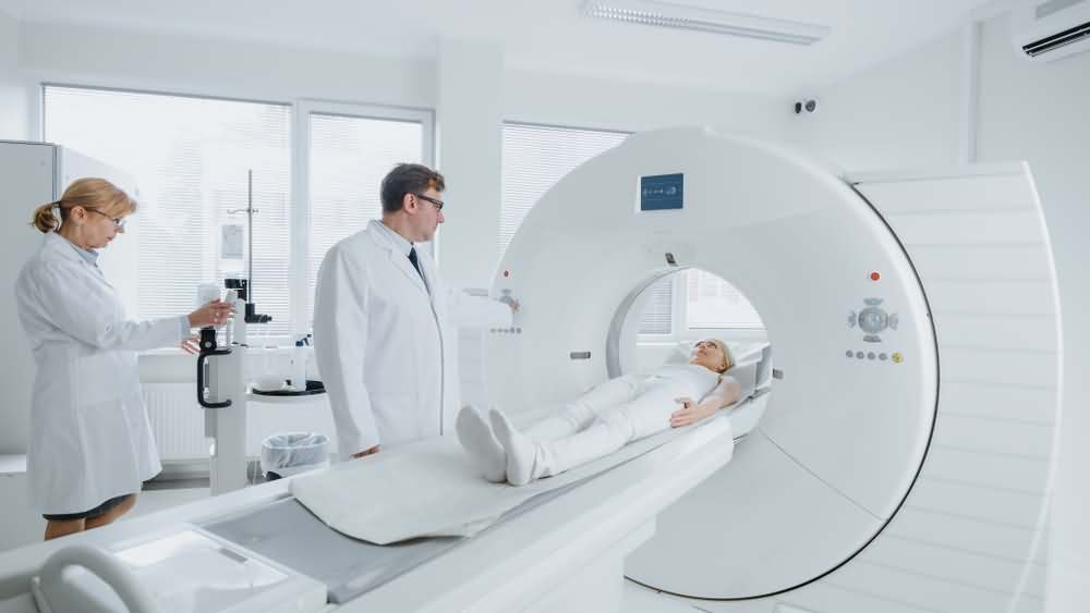
In addition to lab studies, many imaging modalities are used to diagnose jaundice. For example:
- Abdominal ultrasound: Abdominal ultrasound is one of the most commonly done investigations. It is usually requested when an obstructive jaundice is suspected. It can detect the presence of gallstones and may detect if one is lodged in the common bile duct.
- CT: A computed tomography is the best imaging test to detect tumors and their extent. It is done if there is a malignant cause suspected and can detect tumors of the liver and pancreas head. A modification of the test is done by contrast injection. This is especially important in liver tumors.
- MRCP: MRCP is an imaging study that uses magnetic waves to visualize the biliary tract. It can detect stones of the bile canal or if there is a tumor.
The above tests can easily differentiate between gallstones and tumors as causes of obstructive jaundice. Additionally, if the liver is related to the jaundice and the cause is unknown, liver biopsy may be done. It involves sampling a small amount of tissue from the liver to examine under the microscope and detect abnormalities of the liver cells. It can also be done to the pancreas if a tumor is suspected.