Skip to content
How is it performed?

The first step of paracentesis procedures is usually examining the patient through an ultrasound scan. Besides confirming ascites, an ultrasound scan will also provide clues about the amount of fluid in the abdominal cavity, the presence of peritoneal adhesions, and the location of this fluid. After locating the liquid, it is possible to plan for the best site to perform the procedure. This is particularly useful in patients with a smaller volume of fluid.
After addressing this matter and locating the appropriate site for the procedure, and after informed consent has been obtained, the equipment is assembled, and the process starts:
The Process
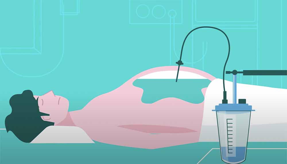
- The patient will be positioned comfortably depending on the puncture site and the liquid’s location. Supine positions are preferred in case of large fluid volume, and lateral decubitus is used in patients with a smaller fluid volume.
- The doctor will gently tap on the abdomen to evaluate the exact position of the fluid. Another option would be guiding the procedure through an ultrasound. The puncture site is then chosen. It can be found in the lower quadrant or the midline. The exact point depends on the patient.
- The skin is then prepared for the puncture and thoroughly cleaned using alcohol solutions. A sterile field is placed around the area, and patients will also receive a local anesthetic to reduce the discomfort during the procedure.
- After the local anesthetic has made effect, the paracentesis needle is fixed and introduced in the puncture site at a perpendicular angle. The doctor will be aspirating as he advances through the soft tissue until he feels how the pressure gives, which is the first sign he is now in the abdominal cavity. The confirmation sign comes when the fluid starts to pass through the syringe.
- The aspirate starts, and it is usually spaced every 50 mL.
- After aspirating the fluid, the needle is swiftly removed while still aspirating. Pressure is applied for short periods in the area, and a bandage is applied.