The Natural Course of the Disease
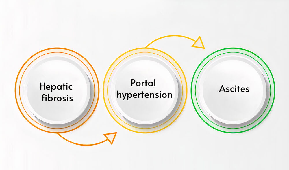
Cirrhosis results from a series of changes to the liver tissue and one of them are hepatic fibrosis. As noted above, hepatic fibrosis is reversible as opposed to cirrhosis. It is an alteration of the components that make up the extracellular matrix of the liver. There’s an excess of type I, type III, and type IV collagen fibers, as well as proteoglycans and glycoproteins. This happens because stellate cells in the liver, which usually create various substances for the extracellular matrix, start synthesizing too much collagen. They are triggered to do this by multiple factors, usually a liver injury, inflammatory cytokines, or toxic substances in the liver tissue.
From this stage, the liver tissue starts undergoing changes that will ultimately lead to portal hypertension when fibrosis transforms into cirrhosis. There’s a deposition of collagen in the space left between the hepatocytes, and the body responds to this excess by creating more blood vessels between the hepatic sinusoids. The stellate cells behave like muscles and create a constriction of the sinusoids simultaneously, and all of this slowly builds up into portal hypertension.
As the cirrhotic liver undergoes fibrosis, it soon develops portal hypertension. This complication results from too much blood entering the liver and a rising resistance against this blood flow. The blood flow increases because there’s an increase in the cardiac output of blood, probably because of a change in nitric oxide levels. There’s also a dilation in the splanchnic arteries that contributes to the process. As noted above, the rising resistance is due to changes in the hepatic sinusoids and stellate cells’ contractile behavior. With too much blood coming in and resistance against blood flow, the liquid is forced out of the blood vessels, in edema and ascites.
Ascites is the most common finding in patients with portal hypertension. It is transudative ascites instead of exudative ascites (found in malignant tumors and other causes of ascites). The difference is that the former has lower protein levels than the latter. The process of ascites formation can be explained by Starling forces that cause liquid diffusion to the extracellular space. From the extracellular space, the excess liquid goes through the lymphatic vessels. In normal cases, these lymph vessels would take care of it and return the liquid to the blood. Still, in portal hypertension, lymph production is more than the lymph vessels can handle, and the fluid escapes and collects into the abdominal cavity.