Skip to content
Diagnosis and Monitoring

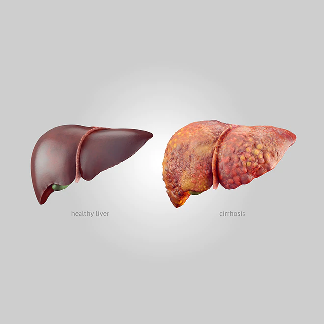
The diagnosis of cirrhosis requires imaging studies as the first confirmation step after considering the patient’s signs and symptoms. In ultrasound studies, it looks nodular instead of smooth, and the organ starts to shrink. The architecture of the liver is irregular and loses its anatomical shape. Similar findings are evident in other imaging studies such as CT scan and MRI.
In diagnosing cirrhosis, it is also essential to rule out other complications such as portal hypertension and hepatorenal syndrome.
To diagnose portal hypertension in cirrhosis, an angiography is required. A catheter is introduced through the femoral or jugular vein and directed into the hepatic vein to measure its pressure. Hepatorenal syndrome is easier to diagnose by using creatinine clearance and measuring creatinine levels in the blood, urine osmolality, and urine sodium levels.
A high number of patients develop ascites along with portal hypertension. In cases of liquid retention in the abdominal cavity, doctors may decide to perform a paracentesis. This exam extracts a sample of liquid from the abdominal cavity to evaluate where it comes from. This exam is useful to rule out other causes of ascites.
Ammonia levels in the blood are also important to measure, especially when diagnosing hepatic encephalopathy. It is routinely done in patients with known cirrhosis or risk factors for cirrhosis who come to the emergency room with seizures, altered mental status, and other signs and symptoms listed above. Sometimes MRI scans and CT scans will be necessary to rule out intracranial lesions.
Given the variable course of cirrhosis, it is always necessary to obtain a score number to evaluate each patient’s severity. For this purpose, the Child-Turcotte-Pugh scoring system combines different variables such as bilirubin levels, albumin in the blood, prothrombin time in seconds, and the presence of ascites and encephalopathy.
After diagnosing and treating patients, and even when they do feel fine, monitoring is vital to evaluate the progression of the disease and prevent further complications. Patients should undergo surveillance for esophageal varices with a full endoscopy every two years and hepatocellular carcinoma through ultrasound scans every 6 months.