
Liver structure and basic anatomy

In its position, the liver has a very complex microscopic structure and a somewhat simple macroscopic structure. In other words, it is a simple organ in plain sight, but under the microscope, it is very complex.
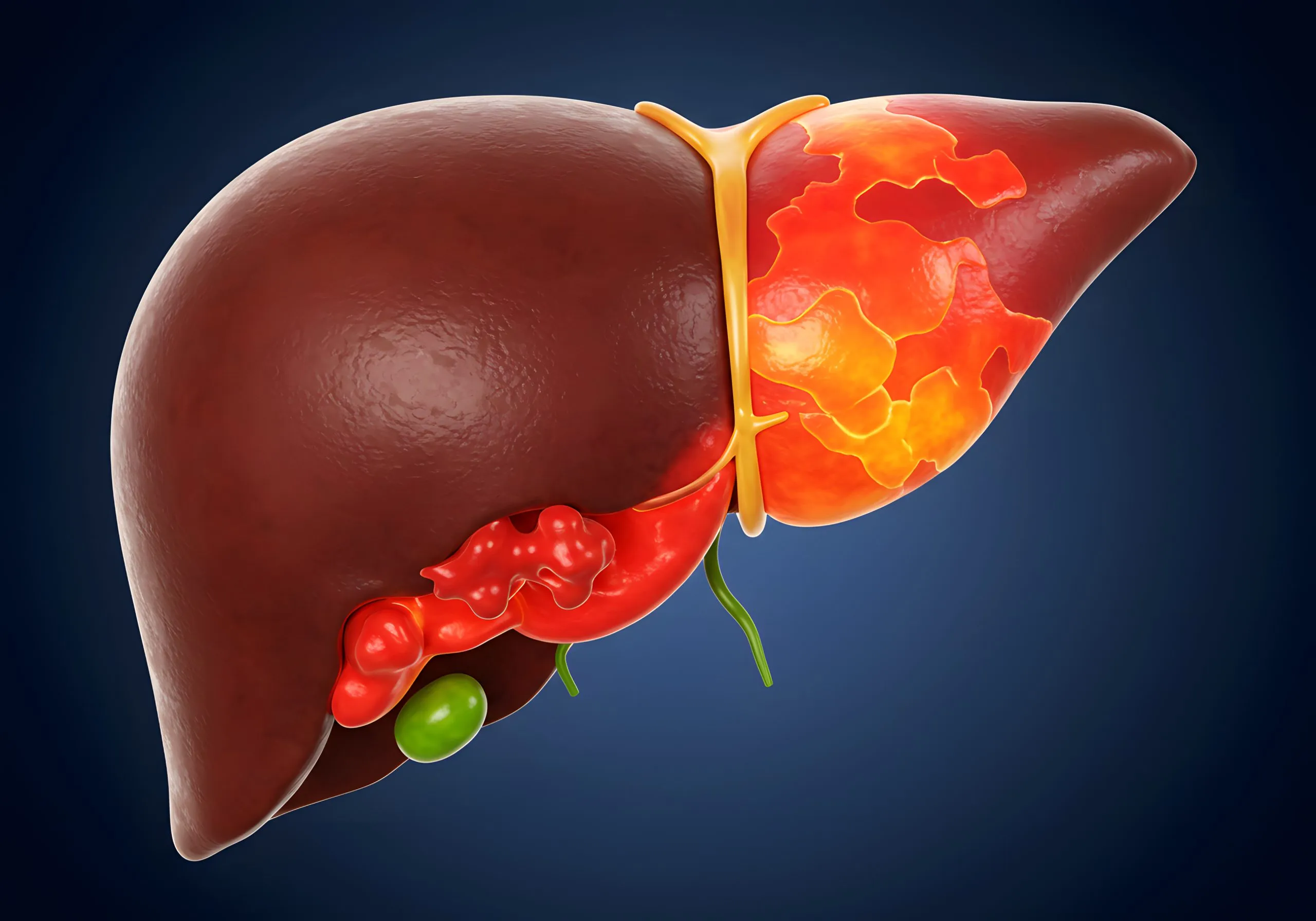
In plain sight, you will see a very smooth organ divided into two sides by a lighter structure called falciform ligament. Thus, we have a huge right lobe and a smaller left lobe. In the middle of the organ, you will also see the gallbladder, and the other structure attached to the liver is the inferior vena cava, which comes directly from the heart and serves the organ as an anchor.
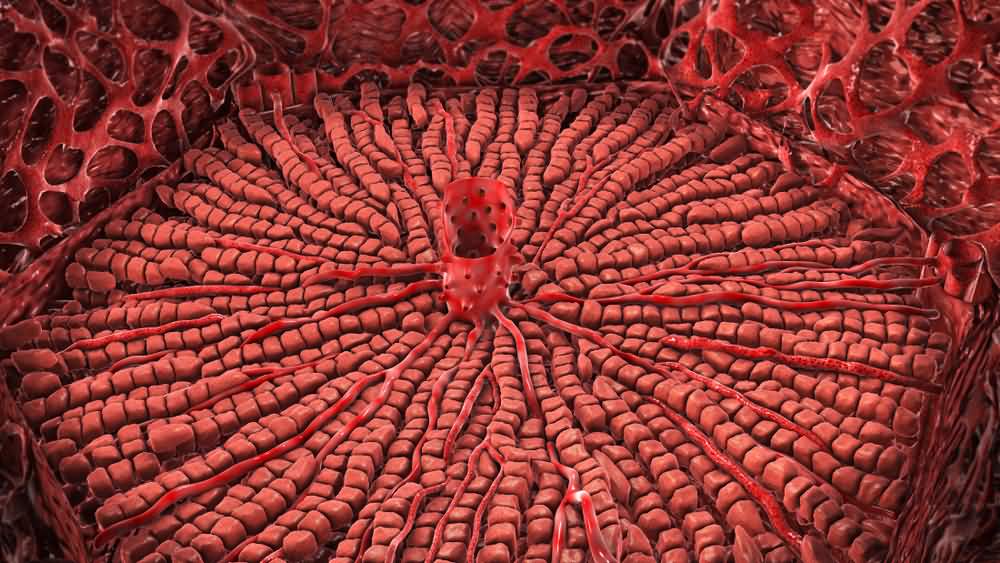
The inner structure of the liver is much more complex when you see it under a microscope. It is formed by cells called hepatocytes. These cells are polygonal in shape and are arranged in sheets of cells around a central vein. So, there’s a central vein, hepatocytes all around, and then another vein surrounded by hepatocytes. This structure of veins and hepatocytes repeats itself all over the organ, called the hepatic lobule. Each hepatic lobule has a central vein, many hepatocytes all around, and a structure known as a portal triad in the corner of the hepatic lobule. Each portal triad contains one small bile duct, one branch of the hepatic artery, and one branch of the hepatic portal vein.
It may seem overwhelming at first, but the structure of the hepatic lobule is easier to understand in a diagram. Once you see it visually, you will get all of the structure of the liver, which copies the same pattern throughout the organ.